


Aseptic bone necrosis
This is a condition in which bone cells die due to insufficient blood supply. This can occur as a result of injury, alcohol abuse, prolonged use of corticosteroids, infections or systemic diseases.
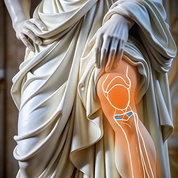
Aseptic necrosis affects bones with a high content of spongy matter: the head of the femur, knee joint, shoulder joint and wrist. The bone begins to break down, which can lead to pain, limited mobility and arthritis.
The symptoms of aseptic necrosis depend on the stage of the disease and the affected area. In the early stages, patients may experience mild pain or discomfort, which increases with physical exertion. As the necrosis progresses, the pain becomes constant, swelling and limitation of joint mobility occur. In advanced cases, bone collapse may occur, which leads to serious deformities and loss of joint function. Aseptic necrosis: what to do? Surgical treatment is indicated in case of ineffectiveness of conservative methods. Surgical intervention can improve blood supply, slow the progression of necrosis and restore joint function. Surgical treatment is indicated in case of ineffectiveness of conservative methods. Surgical intervention can improve blood supply, slow the progression of necrosis and restore joint function.
A general blood and urine test. Coagulogram for the assessment of blood clotting. X-ray, MRI or CT scan to examine the affected area. Consultation with an anesthesiologist to choose the optimal method of anesthesia. Examination and consultation with a therapist for an overall assessment of the patient's health.
In the early stages of the disease, conservative treatment is prescribed: taking painkillers, limiting physical activity, physiotherapy to improve blood flow and strengthen the muscles around the affected bone. When the destruction of bone tissue becomes pronounced, surgical treatment begins. The surgeon can perform decompression surgery to reduce intraosseous pressure, bone marrow or bone marrow cell transplantation, and in case of severe bone damage, endoprosthetics.
X-ray equipment for intraoperative monitoring. A set of metal clamps (plates, screws, pins) to stabilize bone fragments.
After starting treatment, it is important to carry out regular rehabilitation procedures to improve mobility and strength in the affected area. Rehabilitation may include exercises, hydrotherapy, and individual sessions with a physiotherapist. The duration and intensity of rehabilitation measures depend on the general state of the patient's health and the stage of the disease.
Benefits
Restoration of blood supply
Improving blood supply helps to stop necrosis and promotes the restoration of bone cells.
Pain reduction
Relieving symptoms allows patients to return to daily activities and improves their quality of life.
Preventing the progression of the disease
Treatment prevents bone destruction and the need for more radical surgery.
Frequently Asked Questions
What symptoms indicate aseptic bone necrosis?
Is it possible to treat aseptic necrosis with conservative methods?
What methods of prevention of aseptic necrosis exist?
Didn't find an answer to your question?
You can describe your problem in detail and ask a question to the doctor. He will answer you and help you find a solution
Врачи
Смотреть всех врачей
Orthopedic Trauma Surgeon
Useful information
Olecranon fractures
Olecranon has little or no muscle or other soft tissue protection, making it vulnerable to damage from falls and impacts. The olecranon is the most prominent of the bones forming the elbow joint, specifically the ulna. It attaches to the triceps and, along with the humerus, constitutes the elbow joint, which is responsible for bending and extending the arm. Olecranon fractures vary in complexity. They can be simple, with no displacement of bone fragments, or complex, with damage to the articular surface of the ulna and displacement of fragments by triceps traction. Such fractures cause a loss of extension in the elbow joint, resulting from the disruption of the triceps' connection with the forearm.
Forearm fractures
The forearm is composed of two bones, the radius and ulna, which are connected by a firm interosseous membrane and ligaments at the elbow and wrist joints. Due to their intricate structure, defined by numerous curvatures, these bones constitute five joints. The forearm enables flexion and extension at the elbow and wrist joints, as well as the rotation of the hand (pronation and supination) through the movement of the radius around the ulna.
Rotator cuff rupture
An injury characterized by damage to the tendons that surround the shoulder joint and ensure its mobility.
Impeachment-shoulder joint syndrome
Impingement syndrome (subacromial syndrome) is a condition in which the tendons of the rotator cuff of the shoulder are pinched between the acromion and the head of the humerus, which causes pain and limited movement in the shoulder joint.
Instability of the shoulder joint
A pathological condition in which the head of the humerus is displaced relative to the articular cavity of the scapula.
Soft tissue infections
Acute inflammatory processes that occur in subcutaneous tissue, muscles and other soft tissues. They can manifest as abscesses (localized purulent inflammation), phlegmon (diffuse inflammation) or infected wounds.
Similar referral activities
Arthroscopy of the ankle joint
Ankle arthroscopy is a minimally invasive surgical procedure used to diagnose and treat various diseases and injuries of the ankle joint.
Arthroscopy of the knee joint
Knee arthroscopy is a minimally invasive surgical procedure for the diagnosis and treatment of injuries and diseases of the knee joint. It allows examining the joint for damage and eliminating the identified defects.
Arthroscopy of the elbow joint
Arthroscopy of the elbow joint is a minimally invasive surgical intervention that allows for accurate diagnosis and simultaneous treatment of joint injuries.
Arthrodesis of the joints of the fingers of the hand
The destruction of the joints of the fingers of the hand is accompanied by pronounced pain and impaired functions. Arthrodesis is a surgical intervention in which the affected joint is completely immobilized, which relieves pain and progression of inflammation.
Arthroscopic revision of the cystic joint
The condition of the wrist joints determines the functioning of the hand. Arthroscopic revision is a minimally invasive diagnostic procedure that assesses the condition of the joint tissues, which is necessary for planning subsequent treatment.
Arthroscopy of the shoulder joint
Arthroscopy of the shoulder joint is a minimally invasive surgical procedure designed to diagnose and treat various diseases and injuries of the shoulder joint.
News & Media
On April 4 and 5, 2025, the IX ASTAOR International Congress was held — a large-scale meeting of specialists in the field of sports traumatology, orthopedics, arthroscopy and rehabilitation. Doctors, nurses, rehabilitologists and other professionals from different cities of Russia and the world came to share their practical experience and discuss current injury treatment technologies.
Olymp Clinic MARS extends beyond providing high-quality medical care to serve as a comprehensive scientific and educational platform. The clinic will host training events for students, residents, and postgraduates, while physicians will have the opportunity to enhance their qualifications.
An interregional scientific and practical conference, entitled "Advanced Arthroscopic Treatment in Traumatology and Orthopedics," was held in Voronezh on November 1-2. The event convened over 120 specialists from across Russia, including traumatologists, orthopedists, and rehabilitation specialists.
The first manned spacecraft in almost a decade has been successfully launched in the United States. According to many experts, the launch of the world's first private spacecraft marks a new era in space exploration.
A bone fracture is a common injury that almost everyone has been through. Although human bones are very strong, if the external force is too great, they can break like a plastic ruler when it is bent too much. In the article we will tell you how to act in case of a fracture.
Summer is a time for walking, outdoor activities and sports. The sun and warm weather encourage us to spend more time outside and enjoy nature. But the more we move, the higher the risk of injury becomes. It is not surprising that bruises and dislocations become frequent companions of summer leisure. Of course, in case of serious injuries such as fractures, it is necessary to consult a doctor immediately. But what to do with less significant injuries — minor bruises, sprains with slight swelling and pain? In such cases, you can help yourself and others on your own, knowing the basics of first aid. In this article, we will tell you how to cope with injuries sustained during summer activities and quickly return to your usual lifestyle.
How to reach

How to get
From the Belorusskaya metro station of the Zamoskvoretskaya line - exit 4 After exiting the subway, walk through the pedestrian tunnel and climb the stairs. Move towards the railway tracks, go down the stairs immediately after them and walk along the house, then turn right onto 1st Yamskoye Pole Street. At the turn to 3rd Yamsky Pole Street, cross the road at the pedestrian crossing and continue along 1st Yamsky Field Street, after a few buildings on the left you will see Olympus Clinic MARS.
Travel time
9 minutes
Landmark
Olympus Clinic MARS sign
How to get
From the Belorusskaya metro station of the Ring line - exit 2. After exiting the subway, turn left and walk to the pedestrian crossing. Cross the road through two pedestrian crossings and move along the Tverskoy overpass. Go down the stairs immediately after the railway tracks, walk along the house, then turn right onto 1st Yamskoye Pole Street. At the turn to 3rd Yamsky Pole Street, cross the road at the pedestrian crossing and continue along 1st Yamsky Field Street, after a few buildings on the left you will see Olympus Clinic MARS
Travel time
11 minutes
Landmark
Olympus Clinic MARS sign
From the metro station "Tsvetnoy Bulvar"
1 exit to the city, then left to the Garden Ring, at the crossing to the right, crossing the boulevard, one more crossing and at the traffic light to the left. The Olymp Clinic building is located overlooking the Garden Ring to the right of the crossing. Travel time is approximately 9 minutes. Landmark - sign Olymp Clini
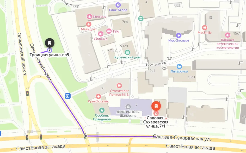
From the metro station "Sukharevskaya"
Exit 3 from the metro and 640 meters straight ahead, the clinic will be on the right. Landmark - sign Olymp Clinic
Parking lot map
Exit 3 from the metro and 640 meters straight ahead, the clinic will be on the right. Landmark - sign Olymp Clinic
From Sokol metro station
The last car from the center: follow the signs for Exit 5. From the glass doors to the right and go to the end of the passage. Exit to the city by the steps to the left. After exiting the crossing to the street, go straight along Leningradsky Prospekt to the intersection with Chapaevsky Lane. Next, turn right (onto Chapaevsky Lane) and walk to the Triumph Palace residential complex. Entrance to the territory: through checkpoint No. 1, opposite the Vkusville store, you will need to present your passport. After passing through the checkpoint, go up the stairs to the fountain, opposite it you will see our clinic.
Travel time
10-12 minutes
From the Airport metro station
The first car from the center: follow the Exit 2-3 signs. Turn left out of the glass doors and walk to the end of the passage. After exiting the crossing to the street, go straight along Leningradsky Prospekt to the intersection with Chapaevsky Lane. Next, turn left (onto Chapaevsky Lane) and walk to the Triumph Palace residential complex. Entrance to the territory: through checkpoint No. 1, opposite the Vkusville store, you will need to present your passport. After passing through the checkpoint, go up the stairs to the fountain, opposite it you will see our clinic.
Travel time
12-15 minutes
How to get
Entry to the territory is prohibited, but there are free city parking lots around the Triumph Palace residential complex, where you can easily find a place for your car. Free parking area:

How to get
From the Belorusskaya metro station of the Zamoskvoretskaya line - exit 4 After exiting the subway, walk through the pedestrian tunnel and climb the stairs. Move towards the railway tracks, go down the stairs immediately after them and walk along the house, then turn right onto 1st Yamskoye Pole Street. At the turn to 3rd Yamsky Pole Street, cross the road at the pedestrian crossing and continue along 1st Yamsky Field Street, after a few buildings on the left you will see Olympus Clinic MARS.
Travel time
9 minutes
Landmark
Olympus Clinic MARS sign
How to get
From the Belorusskaya metro station of the Ring line - exit 2. After exiting the subway, turn left and walk to the pedestrian crossing. Cross the road through two pedestrian crossings and move along the Tverskoy overpass. Go down the stairs immediately after the railway tracks, walk along the house, then turn right onto 1st Yamskoye Pole Street. At the turn to 3rd Yamsky Pole Street, cross the road at the pedestrian crossing and continue along 1st Yamsky Field Street, after a few buildings on the left you will see Olympus Clinic MARS
Travel time
11 minutes
Landmark
Olympus Clinic MARS sign