


Ultrasound of the thyroid gland
A diagnostic method that allows you to evaluate the structure, size and blood supply of the thyroid gland, parathyroid glands and regional lymph nodes. It is indispensable in the diagnosis of diseases such as thyroiditis, thyrotoxicosis, nodular goiter and tumor processes.
The thyroid gland is the main organ of the endocrine system that regulates metabolism and hormonal balance in the body. Disturbances in her work can lead to fluctuations in body weight, increased fatigue, tachycardia and sleep disorders.
The study allows us to see diffuse tissue changes caused by inflammatory diseases. It can be used to detect nodular formations — cysts, adenomas and other tumors, to assess their size and structure. Ultrasound is also recommended for patients with a hereditary predisposition to thyroid diseases, and those who live in regions with iodine deficiency.
Ultrasound of the thyroid gland does not require special training. It is enough for the patient to remove clothes and jewelry from the area under study. Additional examinations may be prescribed, depending on the symptoms and the diagnosis.
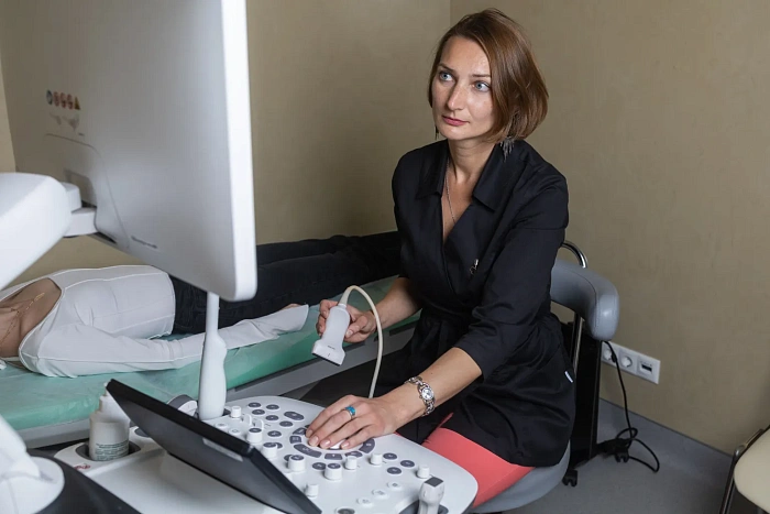
The patient is positioned on his back. A gel is applied to the skin, which improves the conductivity of ultrasound. The examination is carried out using an ultrasonic sensor that moves along the surface of the neck. Ultrasonic waves passing through the tissues form an image of the thyroid gland on the monitor, allowing the doctor to assess the structure and blood flow. The procedure takes about 15-20 minutes.
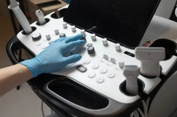
A high-frequency ultrasound machine is used for ultrasound. Modern sensors have high resolution and allow you to get a detailed image of small structures.
After the procedure, the patient can immediately return to normal activities. The results of the study are handed out and sent to the attending physician for analysis and further treatment recommendations.
Benefits
Safety
Ultrasound is safe for all categories of patients, including pregnant women and children.
High precision
It allows you to identify not only structural changes, but also features of blood supply.
Quick diagnosis
The examination takes no more than 20 minutes, and the results are available immediately after the procedure.
The possibility of Dopplerography
Blood flow assessment helps in the diagnosis of diffuse changes in thyroid parenchyma and nodules.
Preparation for the procedure
Ultrasound of the thyroid gland does not require special training. It is enough for the patient to remove clothes and jewelry from the neck and upper chest area.
The course of the procedure
The patient is lying on his back. A gel is applied to the skin, which improves ultrasound conduction. The study is performed using an ultrasound sensor that moves along the surface of the neck. Ultrasound waves passing through the tissues form an image of the thyroid gland on the monitor, which allows the doctor to assess the structure and blood flow. The procedure takes about 15-20 minutes. Based on the ultrasound results, additional examinations and consultations may be scheduled, depending on the identified changes.
The results of the study
After the procedure, the patient can immediately return to normal activities. The results of the study are handed out and sent to the attending physician for analysis and further treatment recommendations.
Frequently Asked Questions
How often do I need to undergo thyroid ultrasound?
Do I need to prepare for a thyroid ultrasound?
Why is thyroid Dopplerography required?
Didn't find an answer to your question?
You can describe your problem in detail and ask a question to the doctor. He will answer you and help you find a solution
Врачи
Смотреть всех врачей
Ultrasound diagnostician, Candidate of Medical Sciences, Higher Qualifying Category Physician. Head of the Functional and Ultrasound Diagnostics department.

Ultrasound Diagnostics Doctor

Ultrasound Diagnostician, Candidate of Medical Sciences, Higher Qualifying Category Physician.

Ultrasound Diagnostics Doctor

Ultrasound Diagnostics Doctor
Similar referral activities
Ultrasound of the joints
A diagnostic method that allows you to assess the condition of soft tissues, tendons, ligaments and cartilage structures of the joint. It is used to detect inflammatory, traumatic and degenerative changes in joints.
Ultrasound of the abdominal cavity
Ultrasound allows you to visualize internal organs such as the liver, gallbladder, pancreas, spleen, kidneys and other structures.
Ultrasound of the gallbladder
A study that allows you to assess the condition and functionality of the gallbladder, as well as identify pathologies such as stones, inflammatory processes and neoplasms.
Ultrasound of the musculoskeletal system
Ultrasound examination, which provides high-precision visualization of muscles, tendons, joints and bone surfaces, allows you to quickly identify injuries and diseases.
Ultrasound of the mammary glands
A diagnostic method that allows you to evaluate the structure of breast tissue and identify pathological changes. Suitable for women of all ages and health conditions.
Ultrasound of soft tissues
A study that allows you to assess the condition of the skin, subcutaneous fat, muscles, tendons, lymph nodes and blood vessels. It helps to identify inflammatory processes, hematomas, tumor formations and traumatic injuries.
Записаться на консультацию
Web-form is not found.