


Computed tomography (CT)
A diagnostic method based on obtaining layered images of the studied area using X-ray radiation. It allows you to get detailed images of internal organs, bones and soft tissues.
CT scan allows you to explore almost any area of the body. The tomograph creates multi-layered images, which allows the doctor to see the smallest details of the area under study.
The procedure is especially effective for the diagnosis of injuries, neoplasms, heart and vascular diseases, pathologies of the respiratory system and bone structures. CT allows you to quickly assess the scale and localization of the pathological process, as well as obtain a three-dimensional image of organs and tissues. CT is used in emergency cases, for example, if a stroke, traumatic brain injury or internal hemorrhage is suspected. This method is also used for primary diagnosis of chest pathologies, suspected cancer or vascular diseases. The procedure can be performed with or without the introduction of a contrast agent. Contrast is used to improve the visibility of blood vessels and soft tissues when tumors or aneurysms are suspected.
It is not recommended to eat 4-6 hours before the contrast study. It is important to inform the doctor about the presence of allergies, kidney failure or diabetes mellitus. The patient must remove all metal objects and jewelry before the procedure.
The patient lies down on a movable table, which moves inside the CT scanner. During the study, the tomograph creates layered X-ray images of the area under study. The data is transferred to a computer, where it is processed and turned into images. If the study is performed with contrast, a contrast agent is injected to the patient before the procedure for better visualization of blood vessels and organs. The procedure takes from 10 to 30 minutes, depending on the area under study. It is important that the patient remains motionless to obtain clear images.
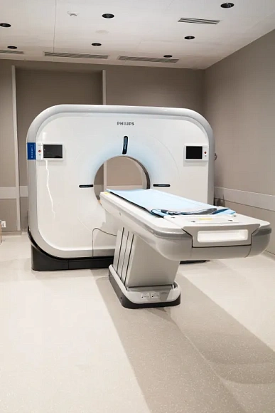
Our clinic uses the Incisive CT CT system to perform computed tomography. The system reduces the radiation load on the patient. The high quality of the images allows you to identify the slightest changes in tissues and organs. Fast data processing allows doctors to quickly make an accurate diagnosis. The system is suitable for examining various areas of the body, including the chest, abdomen, head and spine. The extended diameter of the scanning ring and the quiet operation of the device ensure a comfortable procedure.
After the procedure, the patient can immediately return to normal life. If the CT scan was performed with contrast, it is recommended to drink more water to speed up the removal of the contrast agent from the body.
Benefits
High diagnostic accuracy
CT scans reveal pathologies at an early stage, which significantly improves the prognosis of treatment.
Quick procedure
The study takes only 10-30 minutes, which is important in emergency situations.
Minimum radiation load
Modern devices allow you to reduce the radiation dose without loss of image quality.
The possibility of three-dimensional reconstruction
CT provides not only cross-sectional images, but also a 3D model of organs, which facilitates the diagnosis of complex cases.
Врачи
Смотреть всех врачейSimilar referral activities
Magnetic resonance imaging (MRI)
A diagnostic method that is used to visualize internal organs, tissues, and body systems. The procedure allows you to obtain detailed images of organs without the use of X-rays.
Densitometry
A diagnostic method that allows you to measure bone density and detect osteoporosis in the early stages. The study helps to assess the risk of fractures, determine the level of calcium and the overall structure of bones.
X-ray
A diagnostic method that is used to examine bones, internal organs, and other body structures. X-rays allow you to get an image of internal organs and tissues in different projections.
How to reach

How to get
From the Belorusskaya metro station of the Zamoskvoretskaya line - exit 4 After exiting the subway, walk through the pedestrian tunnel and climb the stairs. Move towards the railway tracks, go down the stairs immediately after them and walk along the house, then turn right onto 1st Yamskoye Pole Street. At the turn to 3rd Yamsky Pole Street, cross the road at the pedestrian crossing and continue along 1st Yamsky Field Street, after a few buildings on the left you will see Olympus Clinic MARS.
Travel time
9 minutes
Landmark
Olympus Clinic MARS sign
How to get
From the Belorusskaya metro station of the Ring line - exit 2. After exiting the subway, turn left and walk to the pedestrian crossing. Cross the road through two pedestrian crossings and move along the Tverskoy overpass. Go down the stairs immediately after the railway tracks, walk along the house, then turn right onto 1st Yamskoye Pole Street. At the turn to 3rd Yamsky Pole Street, cross the road at the pedestrian crossing and continue along 1st Yamsky Field Street, after a few buildings on the left you will see Olympus Clinic MARS
Travel time
11 minutes
Landmark
Olympus Clinic MARS sign
From the metro station "Tsvetnoy Bulvar"
1 exit to the city, then left to the Garden Ring, at the crossing to the right, crossing the boulevard, one more crossing and at the traffic light to the left. The Olymp Clinic building is located overlooking the Garden Ring to the right of the crossing. Travel time is approximately 9 minutes. Landmark - sign Olymp Clini
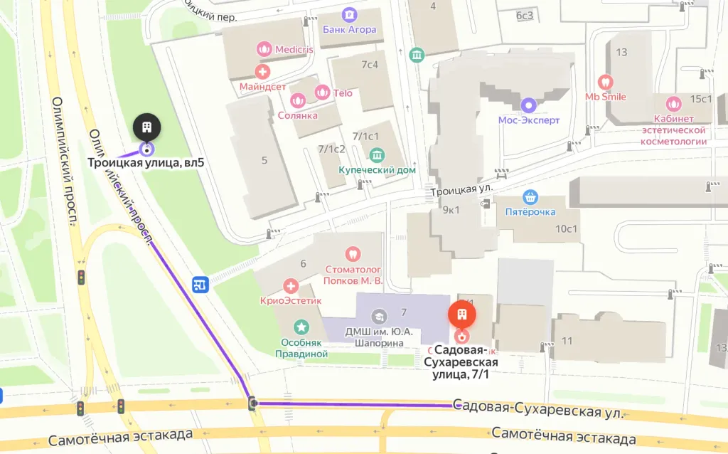
From the metro station "Sukharevskaya"
Exit 3 from the metro and 640 meters straight ahead, the clinic will be on the right. Landmark - sign Olymp Clinic
Parking lot map
Exit 3 from the metro and 640 meters straight ahead, the clinic will be on the right. Landmark - sign Olymp Clinic
From Sokol metro station
The last car from the center: follow the signs for Exit 5. From the glass doors to the right and go to the end of the passage. Exit to the city by the steps to the left. After exiting the crossing to the street, go straight along Leningradsky Prospekt to the intersection with Chapaevsky Lane. Next, turn right (onto Chapaevsky Lane) and walk to the Triumph Palace residential complex. Entrance to the territory: through checkpoint No. 1, opposite the Vkusville store, you will need to present your passport. After passing through the checkpoint, go up the stairs to the fountain, opposite it you will see our clinic.
Travel time
10-12 minutes
From the Airport metro station
The first car from the center: follow the Exit 2-3 signs. Turn left out of the glass doors and walk to the end of the passage. After exiting the crossing to the street, go straight along Leningradsky Prospekt to the intersection with Chapaevsky Lane. Next, turn left (onto Chapaevsky Lane) and walk to the Triumph Palace residential complex. Entrance to the territory: through checkpoint No. 1, opposite the Vkusville store, you will need to present your passport. After passing through the checkpoint, go up the stairs to the fountain, opposite it you will see our clinic.
Travel time
12-15 minutes
How to get
Entry to the territory is prohibited, but there are free city parking lots around the Triumph Palace residential complex, where you can easily find a place for your car. Free parking area:

How to get
From the Belorusskaya metro station of the Zamoskvoretskaya line - exit 4 After exiting the subway, walk through the pedestrian tunnel and climb the stairs. Move towards the railway tracks, go down the stairs immediately after them and walk along the house, then turn right onto 1st Yamskoye Pole Street. At the turn to 3rd Yamsky Pole Street, cross the road at the pedestrian crossing and continue along 1st Yamsky Field Street, after a few buildings on the left you will see Olympus Clinic MARS.
Travel time
9 minutes
Landmark
Olympus Clinic MARS sign
How to get
From the Belorusskaya metro station of the Ring line - exit 2. After exiting the subway, turn left and walk to the pedestrian crossing. Cross the road through two pedestrian crossings and move along the Tverskoy overpass. Go down the stairs immediately after the railway tracks, walk along the house, then turn right onto 1st Yamskoye Pole Street. At the turn to 3rd Yamsky Pole Street, cross the road at the pedestrian crossing and continue along 1st Yamsky Field Street, after a few buildings on the left you will see Olympus Clinic MARS
Travel time
11 minutes
Landmark
Olympus Clinic MARS sign