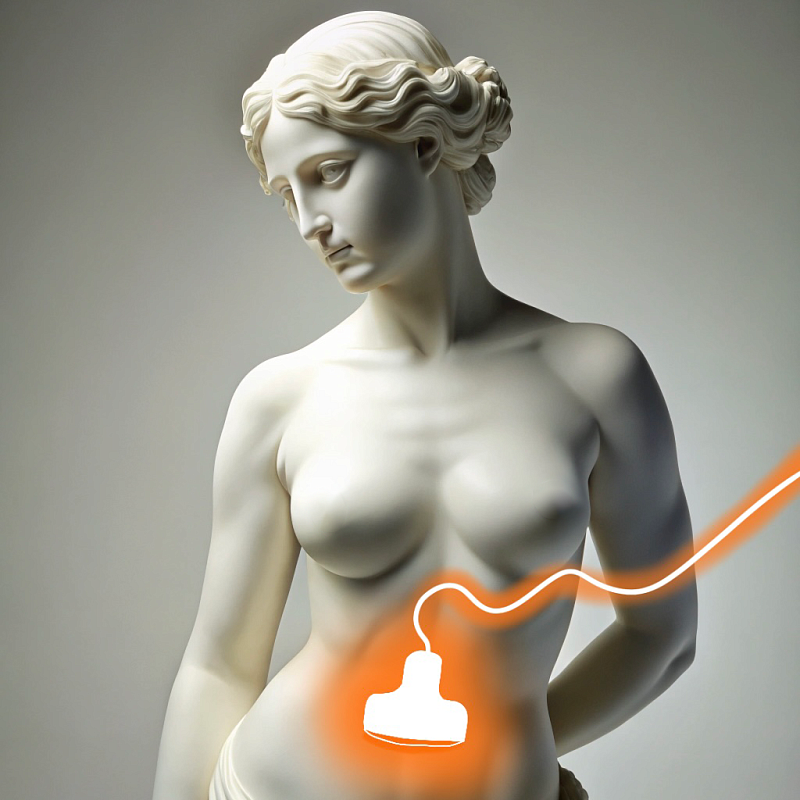
Ultrasound of the pelvic organs
Ultrasound allows you to visualize and assess the condition of the internal organs of the pelvis in women and men. In women, the study includes examination of the uterus, ovaries and fallopian tubes, in men — the prostate gland and seminal vesicles.

In women, the study helps to identify pathologies such as fibroids, cysts, endometriosis, inflammatory processes and tumor formations. In men, the procedure allows you to diagnose prostatitis, prostate adenoma and other diseases.
Benefits
Painlessness and safety
Ultrasound of the pelvic organs is a non—invasive and painless procedure.
High information content
The study provides detailed information about the condition of the internal organs of the pelvis.
Various visualization methods
The possibility of transabdominal, transvaginal and transrectal ultrasound allows you to choose the optimal method of examination.
Speed of implementation
The procedure takes a little time, and the results are available immediately after the study.
Preparation for ultrasound of the pelvic organs
An hour before the study, you should drink 1-1.5 liters of water to fill the bladder, which will improve the visualization of organs. For transvaginal ultrasound (in women) and transrectal ultrasound (in men), special training may be required, which the doctor will inform you about in advance.

Recommendations after ultrasound of the pelvic organs
After performing an ultrasound of the pelvic organs, the patient can immediately return to normal business. It is important to follow the doctor's recommendations, especially if pathologies have been identified that require observation or treatment.

Pelvic ultrasound procedure
For transabdominal ultrasound, the doctor applies a special gel to the skin of the abdomen and moves the sensor, assessing the condition of the pelvic organs through the abdominal wall. For transvaginal ultrasound in women, the sensor is inserted into the vagina, which allows you to get a more detailed image of the uterus and ovaries. For transrectal ultrasound in men, the sensor is inserted into the rectum, which makes it possible to examine the prostate gland in detail. The procedure usually takes about 20-30 minutes.

Frequently Asked Questions
How do I prepare for the procedure?
- Drink 2-3 glasses of water an hour before the study.
- The bladder must be full.
Transvaginal ultrasound (in women):
- No special training is required, the bladder can be emptied.
Transrectal ultrasound (in men):
- 1-2 hours before the study, you can make a cleansing enema, on the recommendation of a doctor.
What organs are examined during ultrasound of the small pelvis in women?
Ovaries: The size, shape, and presence of follicles, cysts, neoplasms, inflammation (oophoritis), and polycystic ovary syndrome (PCOS) are evaluated.
Fallopian tubes: Patency is assessed (with a special ultrasound).
Cervix: The length, structure, and presence of neoplasms are assessed (with transvaginal ultrasound).
How long will it take for the results of the study to be ready?
In some cases, especially with complex studies or high doctor workload, the results may be ready in a few hours.
Less often, the ultrasound results may be ready the next day. This may be due to the need for more detailed image analysis or the workload of the clinic.
How often do I need to undergo the procedure for preventive purposes?
Menopausal women can undergo less often – once every 2-3 years.
If there are risk factors (for example, heredity, hormonal disorders), the frequency may be increased.
Men over 40-45 years old are recommended to undergo once a year for the prevention of prostate diseases, especially in the presence of risk factors (heredity, problems with urination).
For men under 40 years of age, if there are no complaints, it is enough to undergo an ultrasound scan of the prostate gland once every 2-3 years.
Indications for ultrasound of the small pelvis in women
Irregular menstruation
Excessive or poor menstruation
Absence of menstruation (amenorrhea)
Painful menstruation (dysmenorrhea)
Intermenstrual bleeding
Lower abdominal pain:
Acute or chronic abdominal pain
Menstrual cycle-related pain
Pain during sexual intercourse
Suspected gynecological diseases:
Uterine fibroids
Endometriosis
Ovarian cysts
Endometrial polyps
Inflammatory diseases of the uterus and appendages (endometritis, salpingitis, oophoritis)
Ectopic pregnancy
Neoplasms of the pelvic organs
Infertility:
Identification of the causes of infertility, assessment of the condition of the ovaries and uterus
Ovulation assessment
Monitoring of folliculogenesis during ovulation stimulation
Monitoring during assisted reproductive technologies (IVF)
Follow-up after gynecological operations:
Healing control
Detection of complications
Dynamic monitoring of identified formations:
Monitoring the size and growth pattern of fibroids, cysts, and polyps
Assessment of the condition of the endometrium:
In case of pathological bleeding
With infertility
Before starting hormone therapy
Pregnancy:
Early pregnancy diagnosis
Exclusion of ectopic pregnancy
Assessment of fetal condition at various stages
Screening studies during pregnancy
Assessment of the placenta and amniotic fluid
Suspected tumors of the pelvic organs:
Detection of neoplasms of the ovaries, uterus, cervix
Other indications:
Suspected pelvic inflammatory diseases
Diagnosis of complications after childbirth or abortion
Assessment of the bladder condition
What gynecological diseases can be detected by ultrasound of the small pelvis:
Ultrasound allows you to determine the size, number and location of myomatous nodes in the uterus.
The structure of fibroids and their effect on neighboring organs can be assessed.
It is also possible to track the dynamics of fibroid growth during treatment.
Endometriosis:
Ultrasound can reveal foci of endometriosis in the uterus (adenomyosis) and ovaries (endometrioid cysts).
Ultrasound allows you to assess the prevalence of endometriosis and its effect on surrounding tissues.
Ovarian cysts:
Ultrasound allows you to determine the size, structure and number of ovarian cysts (follicular, corpus luteum cysts, endometrioid, dermoid, etc.).
Ultrasound helps to differentiate functional cysts from true neoplasms.
It is possible to assess the dynamics of changes in cysts during the observation process.
Polyps of the endometrium and cervix:
Ultrasound allows you to identify polyps in the uterine cavity and cervical canal.
The size and location of the polyps can be estimated.
Endometrial hyperplasia:
Ultrasound allows you to assess the thickness of the endometrium and identify signs of endometrial hyperplasia (thickening), which may be associated with hormonal disorders or the risk of endometrial cancer.
Pelvic inflammatory diseases (endometritis, salpingitis, oophoritis):
Ultrasound can reveal signs of inflammation of the uterus, fallopian tubes, and ovaries, such as thickening of the walls, and the presence of fluid in the pelvis.
Ultrasound can help identify purulent formations (abscesses) in the appendage area.
Ectopic pregnancy:
Ultrasound may reveal a fetal egg outside the uterine cavity, which is a sign of an ectopic pregnancy.
Ultrasound can also help determine the location of an ectopic pregnancy (fallopian tube, ovary, abdominal cavity).
Neoplasms of the uterus and ovaries:
Ultrasound may reveal suspected malignancies of the uterus, ovaries, and cervix, although additional studies (biopsy, histological examination) may be required to confirm the diagnosis.
Ultrasound helps to assess the size, structure, and prevalence of tumors.
Abnormalities of uterine development:
Ultrasound can reveal abnormalities of the uterus, such as a bicorn uterus, saddle uterus, and others.
The condition of the endometrium during various phases of the menstrual cycle:
Ultrasound allows you to assess the thickness and structure of the endometrium depending on the phase of the menstrual cycle, which may be important in the diagnosis of infertility.
Folliculogenesis monitoring:
Ultrasound is used to monitor the growth of follicles in the ovaries, which is important in the treatment of infertility and ovulation stimulation.
Didn't find an answer to your question?
You can describe your problem in detail and ask a question to the doctor. He will answer you and help you find a solution
Врачи
Смотреть всех врачейUltrasound diagnostician, Candidate of Medical Sciences, Higher Qualifying Category Physician. Head of the Functional and Ultrasound Diagnostics department.
Ultrasound Diagnostics Doctor
Indications and contraindications
Indications
Pain and discomfort in the pelvic area
Pain in the lower abdomen, which may be associated with diseases of the internal organs of the pelvis.
Menstrual disorders in women
The study helps to identify the causes of cycle disorders such as fibroids, cysts and endometriosis.
Problems with urination in men
With difficulty urinating, pain and other symptoms indicating pathology of the prostate gland.
Pregnancy planning and monitoring
To assess the condition of the uterus and ovaries during pregnancy planning and during prenatal follow-up.
Expected effect
Accurate diagnosis
Ultrasound allows you to identify and evaluate diseases of the pelvic organs.
Monitoring of chronic diseases
Monitoring the condition of patients with chronic diseases of the pelvic organs.
Identification of hidden pathologies
Detection of diseases in the early stages, which allows you to start timely treatment.
Assessment of the state of reproductive organs
In women, the study helps in planning pregnancy and monitoring its course, in men — in assessing the condition of the prostate gland and seminal vesicles.
Similar referral activities
Ultrasound of the joints
A diagnostic method that allows you to assess the condition of soft tissues, tendons, ligaments and cartilage structures of the joint. It is used to detect inflammatory, traumatic and degenerative changes in joints.
Ultrasound of the abdominal cavity
Ultrasound allows you to visualize internal organs such as the liver, gallbladder, pancreas, spleen, kidneys and other structures.
Ultrasound of the gallbladder
A study that allows you to assess the condition and functionality of the gallbladder, as well as identify pathologies such as stones, inflammatory processes and neoplasms.
Ultrasound of the musculoskeletal system
Ultrasound examination, which provides high-precision visualization of muscles, tendons, joints and bone surfaces, allows you to quickly identify injuries and diseases.
Ultrasound of the mammary glands
A diagnostic method that allows you to evaluate the structure of breast tissue and identify pathological changes. Suitable for women of all ages and health conditions.
Ultrasound of soft tissues
A study that allows you to assess the condition of the skin, subcutaneous fat, muscles, tendons, lymph nodes and blood vessels. It helps to identify inflammatory processes, hematomas, tumor formations and traumatic injuries.

