


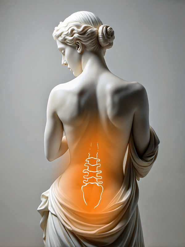
Scoliosis and kyphosis spine surgery (Spine straightening)
Surgical treatment for scoliosis and kyphosis is performed when the spinal curvature progresses and causes pain or impaired posture, or when the patient has a pronounced cosmetic defect. The procedure aims to restore the correct spinal alignment, address cosmetic deformities, and stabilize the spine.
The surgery is classified according to its degree of complexity depending on the severity of the curvature, the extent of the intervention, and associated risk.
Before the operation, an extensive examination is carried out. This includes blood and urine tests, spinal MRI and CT scans, and an ECG. These tests evaluate the extent of the deformity, identify any contraindications, and determine the appropriate anesthesia. The patient is examined by a spine specialist, a general practitioner, and an anesthesiologist. If necessary, functional X-rays are performed. Based on the results, the extent of the correction and the type of metal structure are planned.
The procedure is performed under general anesthesia. First, the surgeon accesses the spine. Then, screws are inserted into the supporting vertebrae. Next, the curvature is corrected by fixing the spine in its new position with rods. Complex cases may require osteotomy, which involves removing parts of the bone to facilitate straightening. Complicated cases require a combined approach or multi-stage intervention. All actions are controlled using X-ray navigation and body function monitoring systems to ensure accurate and safe correction of the spinal axis.
The surgery requires advanced neurosurgical equipment, such as X-ray imaging, navigation systems, and internal organ function monitoring systems. We also use stabilization systems made of titanium from leading international manufacturers. These systems are designed for permanent installation and will keep your spine stable, even under heavy loads.
The early postoperative period is spent in the hospital. On the first day, the patient begins walking independently and receives pain relief. Typically, patients need to stay in the hospital for 5-6 days after surgery. During this time, physical therapists work with patients to speed up their recovery. Wearing a corset after surgery is not required. After discharge, a recovery program, physical and exercise therapy are prescribed. Physical activity is restricted for 3-6 months. Regular checkups and X-ray examinations are conducted to assess the stability of the structure and the progress of fusion.
Benefits
Aesthetic effect
The operation straightens the spine, significantly improving posture and the aesthetic appearance of the body
Improved quality of life
Chronic pain is eliminated and overall well-being is improved
Long-term results
The latest fixation methods stabilize the spine and prevent future deformity progression
Precision
Advanced technology and experienced surgical team ensure accuracy and safety during surgery
Врачи
Смотреть всех врачей
Orthopedic Trauma Surgeon, Vertebrologist

General surgeon, Professor, Doctor of Medical Sciences. Head of the Spine Surgery Department.
Similar referral activities
Endoscopic stenosis decompression
Endoscopic stenosis decompression is a minimally invasive surgical procedure designed to relieve pressure on the spinal cord and nerve roots caused by narrowing of the spinal canal.
Endoscopic discectomy
Endoscopic discectomy is a minimally invasive surgical procedure aimed at removing pathological disc protrusion that puts pressure on the nerve, with minimal impact on the surrounding tissue.
X-ray-guided transforaminal injection (1 zone)
X-ray-guided transforaminal injection provides targeted relief for pain caused by pinched or inflamed nerve roots in the intervertebral foramen area. This method ensures the accurate administration of drugs with minimal trauma.
Goel-Harms fixation
Goel-Harms fixation is a standard decompression and stabilization technique used for compression fractures, spinal stenosis, segmental instability, or spondylolisthesis.
X-ray-guided facet joint injection (1 spinal segment)
X-ray-guided facet joint injection is a minimally invasive procedure aimed at eliminating pain caused by inflammation or degenerative-dystrophic changes in the facet joints of the spine.
X-ray-guided knee or hip joint nerve injection
Knee or hip joint nerve block is an injection procedure that helps relieve pain caused by degenerative, inflammatory, or postoperative changes in the joints.