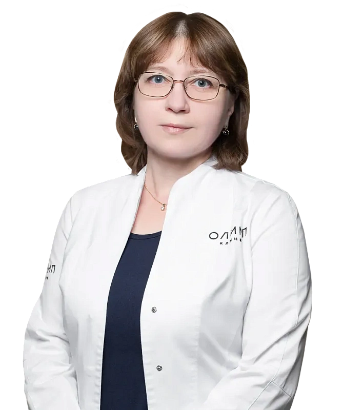
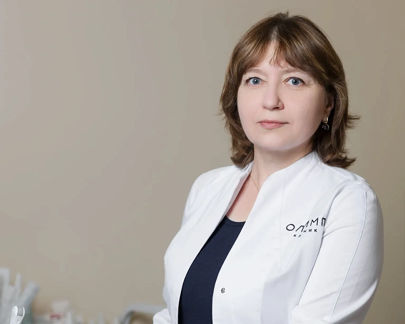
Novikova Inna Valeryevna
Oncologist, surgeon, mammologist, ultrasound diagnostics doctor
Patients
from 18 yearsLanguages
English, German
Inna Valeryevna is a specialist in the diagnosis of breast diseases, ultrasound of the mammary glands, fine needle puncture, biopsy of nodular formations, puncture of cystic formations under ultrasound control.
You're in good hands.
Inna Valeryevna will be able to understand the problem and help each patient.

Education and qualifications
1999
Pediatrics
Russian State Medical University
2001
Therapy
Russian State Medical University
2001-2009
Assistant of the Department of Radiation Diagnostics, Radiation Therapy
Russian State Medical University
2001-2024
Oncologist, ultrasound diagnostics doctor
D.D. Pletnev City Clinical Hospital
2003
Radiology
Russian Medical Academy of Postgraduate Education
2006
Oncology
Russian State Medical University
Take a look at the work of specialists
Frequently Asked Questions
What risk factors can increase the likelihood of breast cancer, and how can I minimize these risks?
Factors affecting the risk of breast cancer can be divided into uncontrolled:
- with increasing age, the risk of breast cancer increases, reaching its maximum
maximum in postmenopause.
- the presence of mutations in genes whose function is related to cell cycle regulation and/or
DNA repair - BRCA1 and BRCA2, p53, PTEN, etc.
and controlled:
- age during the first pregnancy
- number of pregnancies, IVF
- the number of abortions
- breastfeeding
- taking hormonal drugs
- concomitant gynecological pathology, endocrine diseases
- diet and exercise level
- alcohol consumption
- exposure to ionizing radiation, mechanical injuries, operations
- working conditions with occupational hazards
Doctor: Novikova Inna Valeryevna
Does breast ultrasound give a 100% result or may it not detect the presence of pathology?
In the first place among the large arsenal of modern methods for diagnosing breast diseases
remains a clinical examination, consisting of anamnesis collection, examination and
palpation of the mammary glands and regional lymph nodes.
Ultrasound has been used to examine the breast since the mid-70s of the last
century, for more than 50 years. Currently, ultrasound of the mammary glands is widely used to
detect diffuse mastopathy, ductal pathology, cysts, benign nodular
pathology, and malignant tumors.
This is a highly informative, effective, non-invasive and safe method that has its
advantages.:
- the possibility of widespread use in all women, with and without complaints,
for preventive purposes
- safety and information in pregnant and lactating women (for example, during diagnosis
lactostasis, mastitis)
- no radiation exposure
- no special training is required before ultrasound
However, it is worth noting that no diagnostic method, even the most modern one, gives a 100%
result.
The main disadvantages of ultrasound include:
- low information content in case of breast fat involution
- the inability to visualize small non-palpable tumors up to 4-5 mm,
represented by an accumulation of microcalcinates, and against the background of a local severe
restructuring of the structure of the mammary glands.
Ultrasound is not the only, but only one of the methods of comprehensive breast examination.
Mammography, MRI, and other radiation diagnostic methods are used to visualize the breast, in addition to ultrasound examination.
Women over the age of 40 need to perform mammography of both mammary glands in two
projections (Order of the Ministry of Health of the Russian Federation dated January 28, 2021 No. 29N) once every 2 years, in
over the age of 50 – annually.
Doctor: Novikova Inna Valeryevna
When is it better and more correct to be examined by a mammologist: before, during or after menstruation?
A woman of any age should perform monthly breast self-examination, and
if changes are detected, consult a specialist. Healthy women aged 19-39 years
are regularly examined by a mammologist at least once every 2 years.
The ultrasound picture of unchanged mammary glands is variable, in the reproductive period
it primarily depends on the phase of the menstrual cycle. For any breast examination
, the optimal days after the end of menstruation are from the 5th to the 14th day of the month.
Doctor: Novikova Inna Valeryevna
Didn't find an answer to your question?
You can describe your problem in detail and ask a question to the doctor. He will answer you and help you find a solution
Specialists in this department
Find a Specialist
Surgery
Learn more
Experience: 24 года
Novikova Inna Valeryevna
Oncologist, surgeon, mammologist, ultrasound diagnostics doctor


