


Plastic medial holder of the patella
Dislocation of the patella occurs at the moment of twisting in the joint and displacement of the kneecap: falling to the knee, hitting the straightened leg in the knee area. A gentle method of surgical treatment is arthroscopic intervention.
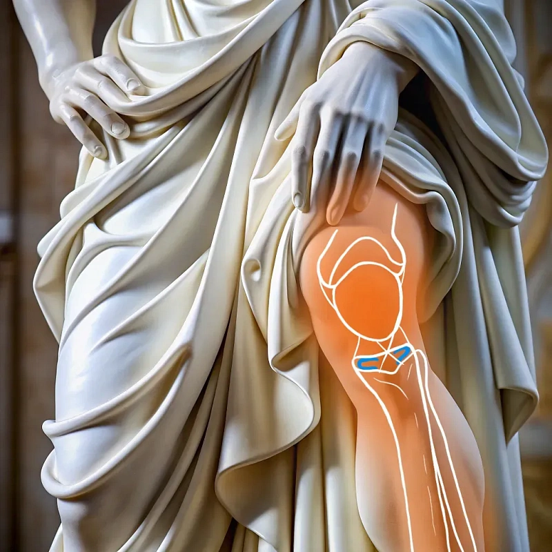
The mechanism of injury occurs as follows: the knee turns inward, the retaining muscle is sharply stretched. As a result, the internal restraint breaks and dislocation occurs. With repeated traumatization, osteoarthritis may develop.
Restoration of the integrity of the ligaments is performed using arthroscopic plastic surgery. During the operation, the patella "returns" to its place, the anatomical integrity of the joint is restored. Plastic surgery of the medial patellar retainer is performed by experienced sports orthopedic traumatologists and surgeons, which guarantees high efficiency of the procedure.
Clinical analysis of blood and urine Radiography of the knee joint Magnetic resonance imaging (MRI) Consultation with a therapist and specialized specialists such as an orthopedic traumatologist and an anesthesiologist
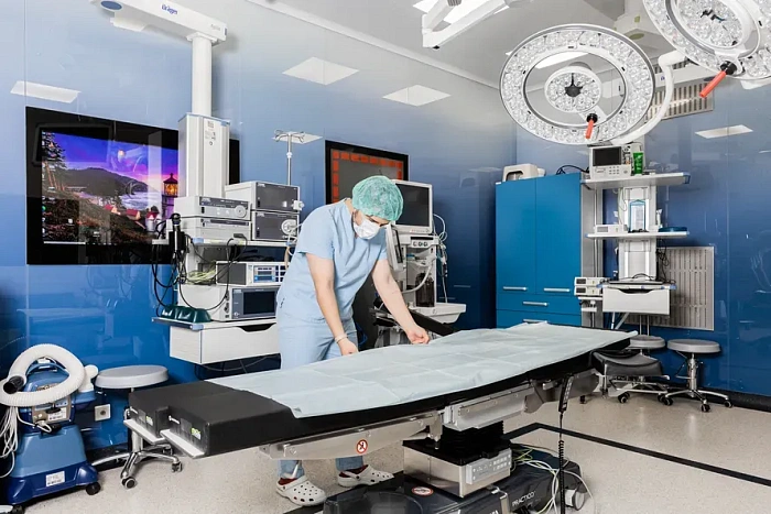
On the day of the operation, the patient is hospitalized in the morning, and laryngeal mask anesthesia is performed. A camera with instruments is inserted into the joint through micro-injections, with the help of which the condition of internal structures is assessed and the necessary manipulations are performed. The surgeon carefully adjusts the medial patellar retainer by strengthening existing ligaments or creating new ones fr om the patient's tissues or surgical materials. After the operation, the patient is transferred to the awakening room, wh ere his condition is monitored by medical staff. The operation lasts an average of 60-90 minutes.
Arthroscopes with high-quality optics Miniature surgical instruments
For the first 3 weeks, the knee joint is fixed in a straight orthosis, and the movement is carried out with the help of crutches. Then therapeutic gymnastics and physiotherapy (magnetotherapy, UVT, kinesiotaping, massage) are performed with a gradual increase in the load on the knee. After 4 weeks, a gradual transition from crutches to a cane begins, and then to walking without support. Full recovery takes 3-4 months.
Benefits
Precise correction
Return to the correct position of the patella.
Minimal invasiveness
The use of low-traumatic surgical techniques.
Fast recovery
Modern methods of treatment and rehabilitation contribute to a quick return to normal life.
Long-term result
Achieving a long-lasting effect of knee stability.
Preparation for plastic surgery of the medial patellar retainer
Before surgery, the patient undergoes a full medical examination, including an MRI or ultrasound of the knee joint, for accurate diagnosis and planning of surgical intervention. It is important to discuss with your doctor all the potential risks and expectations of surgery.
Plastic surgery of the medial patellar retainer
The surgeon makes several small incisions around the knee joint to insert arthroscopic equipment — a thin tube with a camera and a lighting device at the end. During the operation, the surgeon carefully adjusts the medial patellar retainer, strengthening existing ligaments or creating new ones from the patient's tissues or surgical materials.
Recommendations after plastic surgery of the medial patellar retainer
After surgery, the patient may be advised to wear an orthopedic splint to stabilize the knee, as well as perform exercises under the guidance of a physiotherapist to restore strength and mobility of the joint. It is important to avoid intense stress on the knee during the first weeks after surgery.
Frequently Asked Questions
How long does the operation take?
After what time can you return to normal life?
Under what anesthesia is the operation performed?
Didn't find an answer to your question?
You can describe your problem in detail and ask a question to the doctor. He will answer you and help you find a solution
Врачи
Смотреть всех врачей
Doctor of Medical Sciences, Professor. Orthopedic Trauma Surgeon. Chief Doctor, Medical Director.

Doctor of Medical Sciences, Associate Professor at the Department of Traumatology and Orthopedics of the RUDN. Head of the Center for Arthroscopy and minimally invasive surgery of the joints of the upper and lower extremities.
Indications and contraindications
Indications
Injuries to the medial retainer
Ruptures or sprains that require surgery to repair.
Patellar deformities
Anatomical abnormalities that lead to a violation of its function and position.
Chronic instability
Permanent dislocations or subluxations that interfere with normal life activity.
Pain syndrome
Chronic knee pain associated with patellar dysfunction.
Expected effect
Restoring knee stability
Reduction or elimination of dislocations and subluxations of the patella.
Improving joint functionality
Return to sports and daily activities without restrictions.
Reducing discomfort
Relief of knee pain that occurs during movement or exertion.
Prevention of degenerative changes
Slowing down the development of osteoarthritis and other degenerative diseases of the knee joint.
Similar referral activities
Arthroscopy of the ankle joint
Ankle arthroscopy is a minimally invasive surgical procedure used to diagnose and treat various diseases and injuries of the ankle joint.
Arthroscopy of the knee joint
Knee arthroscopy is a minimally invasive surgical procedure for the diagnosis and treatment of injuries and diseases of the knee joint. It allows examining the joint for damage and eliminating the identified defects.
Arthroscopy of the elbow joint
Arthroscopy of the elbow joint is a minimally invasive surgical intervention that allows for accurate diagnosis and simultaneous treatment of joint injuries.
Arthrodesis of the joints of the fingers of the hand
The destruction of the joints of the fingers of the hand is accompanied by pronounced pain and impaired functions. Arthrodesis is a surgical intervention in which the affected joint is completely immobilized, which relieves pain and progression of inflammation.
Arthroscopic revision of the cystic joint
The condition of the wrist joints determines the functioning of the hand. Arthroscopic revision is a minimally invasive diagnostic procedure that assesses the condition of the joint tissues, which is necessary for planning subsequent treatment.
Arthroscopy of the shoulder joint
Arthroscopy of the shoulder joint is a minimally invasive surgical procedure designed to diagnose and treat various diseases and injuries of the shoulder joint.