


Treatment of pelvic bone fractures
Fractures of the pelvic bones sometimes occur in car accidents and falls from a great height. Often a fracture is part of a complex polytrauma with damage to many organs and tissues and requires urgent medical intervention.
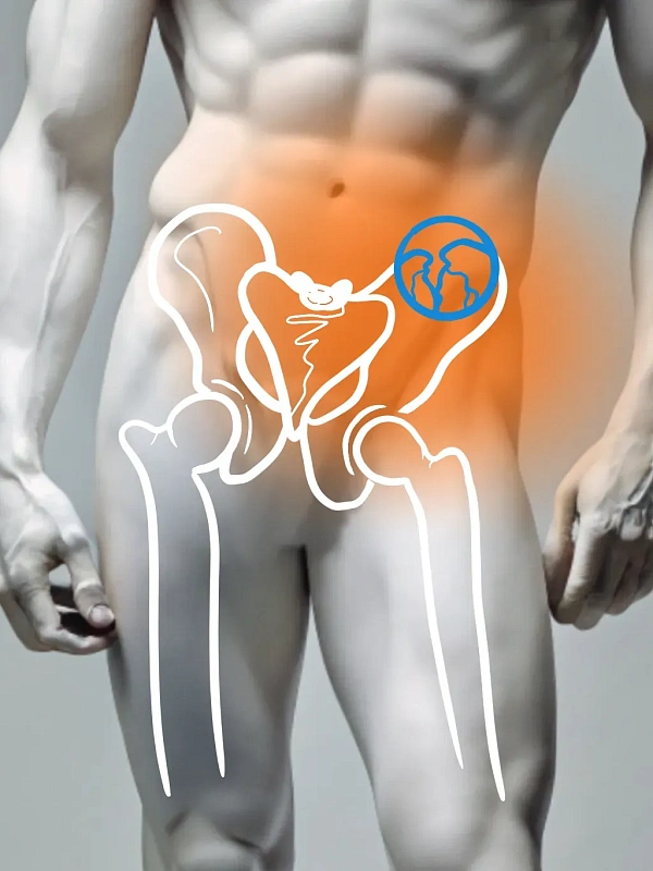
The pelvic bones support the upper body, provide movement and protect internal organs. Stable pelvic bone fractures are usually not displaced and can heal with nonsurgical treatment, while unstable fractures require surgical intervention.
Pelvic fractures are often accompanied by internal organ and blood vessel damage, making them particularly dangerous and requiring immediate medical attention. Major symptoms of pelvic bone fractures include severe pelvis and lower back pain, impaired movement or walking, swelling and bruising. Treatment of pelvic fractures aims to stable the bone, relieve pain and prevent complications. Osteosynthesis allows precise fixation of the bone fragments, which promotes proper fusion and reduces the risk of dislocation.
Common blood and urine tests Coagulogram (blood clotting assessment). X-ray or computerized tomography (CT) scan of the pelvis. Electrocardiogram (ECG). Consultation with an anesthesiologist to choose the best anesthesia option. Examination and consultation with a general practitioner for a general assessment of the patient's health.
Treatment of pelvic bone fractures is based on the type and extent of the fracture. In cases of minor or non-displaced fractures, a conservative approach is used, including anesthesia, blood compensation and fracture immobilization. Displaced, unstable or complex fractures require surgical intervention. Surgical techniques may include open reduction and internal fixation of the bone fragments with steel plates, screws and pins. Some cases require external fixation.
X-ray equipment for intraoperative monitoring and precise positioning of the retainers. Steel retainer set (plates, screws, pins) to fixate bone fragments.
Rehabilitation starts after treatment, to restore mobility, strength and lower body function. The physician may prescribe physiotherapy, exercise therapy and, optionally, massage. An important part of rehabilitation is adapting the patient to new lifestyle changes and minimizing the impact of the injury on daily activities. The rehabilitation program is selected individually and is based on the patient's overall health and the extent of the fracture.
Benefits
Anatomy restoration
Proper joining and securing bone fragments restores pelvis function and mobility.
Pain relief
Treatment helps patients return to regular activity and independent movement.
Preventing complications
Treatment minimizes the risk of chronic pain and pelvic organ dysfunction.
Frequently Asked Questions
Is surgery required for fractured pelvic bones?
How long does rehabilitation take after a pelvic bone fracture?
What symptoms indicate a pelvic bone fracture?
What precautions should be taken after a pelvic bone fracture?
Didn't find an answer to your question?
You can describe your problem in detail and ask a question to the doctor. He will answer you and help you find a solution
Врачи
Смотреть всех врачей
Candidate of Medical Sciences. Orthopedic Trauma Surgeon. Head of the Traumatology Department.
Similar referral activities
Arthroscopy of the ankle joint
Ankle arthroscopy is a minimally invasive surgical procedure used to diagnose and treat various diseases and injuries of the ankle joint.
Arthroscopy of the knee joint
Knee arthroscopy is a minimally invasive surgical procedure for the diagnosis and treatment of injuries and diseases of the knee joint. It allows examining the joint for damage and eliminating the identified defects.
Arthroscopy of the elbow joint
Arthroscopy of the elbow joint is a minimally invasive surgical intervention that allows for accurate diagnosis and simultaneous treatment of joint injuries.
Arthrodesis of the joints of the fingers of the hand
The destruction of the joints of the fingers of the hand is accompanied by pronounced pain and impaired functions. Arthrodesis is a surgical intervention in which the affected joint is completely immobilized, which relieves pain and progression of inflammation.
Arthroscopic revision of the cystic joint
The condition of the wrist joints determines the functioning of the hand. Arthroscopic revision is a minimally invasive diagnostic procedure that assesses the condition of the joint tissues, which is necessary for planning subsequent treatment.
Arthroscopy of the shoulder joint
Arthroscopy of the shoulder joint is a minimally invasive surgical procedure designed to diagnose and treat various diseases and injuries of the shoulder joint.